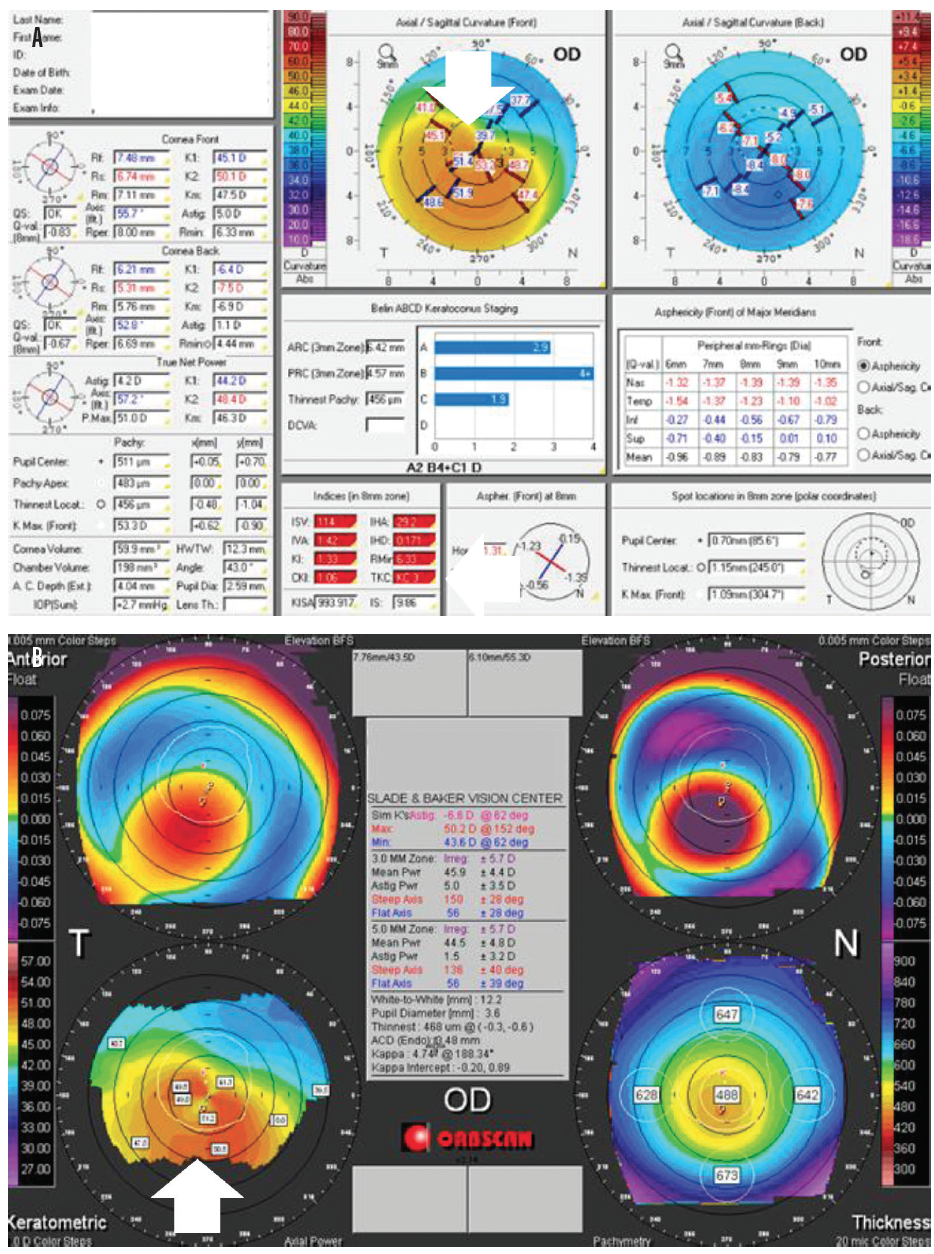

The area superior to the thinned part is ectatic, and the area between the limbus and thinned part is clear, without any scarring, lipid deposition, or vascularization. The band of thinning usually extends between the 4 and 8 o'clock positions and is apart from the limbus by 1–2 mm of the normal cornea. 1 PMD is different from other ectatic corneal disorders by its location. Dr.Pellucid marginal degeneration (PMD) is a non-inflammatory and progressive ectatic corneal disease involving the inferior cornea in a crescentic shape.

Nordan, MD, is a technology consultant for Vision Membrane Technologies, Inc., in San Diego. For now, given the known efficacy of PRK and the grave consequences of ectasia following LASIK, I recommend that surgeons not perform LASIK on corneas that have a central thickness of 500 µm or less preoperative irregular astigmatism. His research is sorely needed and may answer many questions about the risk factors for ectasia after LASIK. Doyle Stulting, MD, PhD, of the Emory Eye Center in Atlanta is conducting an extensive study that will correlate corneal pachymetry, other corneal factors, and LASIK outcomes.

The term forme fruste keratoconus is a misnomer, primarily because the presence of even a small degree of irregular astigmatism generally indicates overt-albeit mild-keratoconus. I define forme fruste ectasia as a condition of the cornea that includes reduced corneal strength and corneal thinning (usually centrally or inferiorly) without irregular astigmatism. This article shares some of my thoughts on forme fruste ectasia. All of the experts agreed that this inferiorly thinning profile was abnormal. If forme fruste keratoconus or forme fruste corneal pellucid marginal degeneration exists, either can cause the inferior cornea to be equal to or thinner than the central cornea. Nonetheless, these areas of thinning can overlap.

Keratoconus and corneal pellucid marginal degeneration are part of the same ectatic disease complex, but they have a different primary location (Figure 1). My interpretation of this small but important sampling is as follows. During this session, I conducted a written poll of the eight panelists for this discussion and discovered some interesting facts (Table 1).
#Pellucid marginal degeneration ectasia update
Schallhorn, MDĭuring the Storm Eye/ASCRS Clinical Update 2007, held from May 31 to June 3, one of the hot topics of the corneal section was attempting to identify the risk factors for ectasia before LASIK. The Effect of PRK and LASIK on Corneal Keratocytesĥ Questions with Steven C. Pearls for Success With Presbyopia-Correcting IOLs Point/Counterpoint: Is Corneal Thickness a Risk Factor for Post-LASIK Ectasia? Combined Surgery in a Uniocular, Mentally Retarded Patient


 0 kommentar(er)
